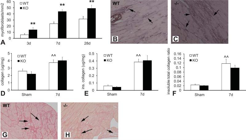
Figure 3.
Although TSP-1 null pressure-overloaded hearts are infiltrated with abundant fibroblasts, collagen content is not significantly increased. A-C Myofibroblasts were identified in the pressure overloaded myocardium as spindle-shaped cells expressing α-SMA located outside the vascular media (arrows). Quantitative analysis demonstrated that myofibroblast density was significantly higher in TSP-1 -/- hearts after 3-28 days of TAC (**p<0.01 vs. WT). Representative images showing α-SMA staining to identify myofibroblasts in WT (B) and TSP-1 null (C) hearts after 7 days of pressure overload are shown. D-F. Cardiac pressure overload was associated with increased total and insoluble collagen content (measured with a hydroxyproline biochemical assay) and a higher ratio of insoluble:total collagen (^^p<0.01 vs. sham). Despite a markedly increased myofibroblast density, collagen content was not significantly higher in TSP-1 null hearts after 7 days of pressure overload (D). The amount of insoluble collagen (E) and the ratio of insoluble:total collagen (F) were comparable between TSP-1 -/- and WT animals. Sirius red staining showed that, while pressure overloaded WT hearts had interstitial deposition of dense collagen (arrows), TSP-1 -/- animals had more extensive cardiomyocyte loss, associated with replacement by a matrix network comprised of loose collagen (arrows).

