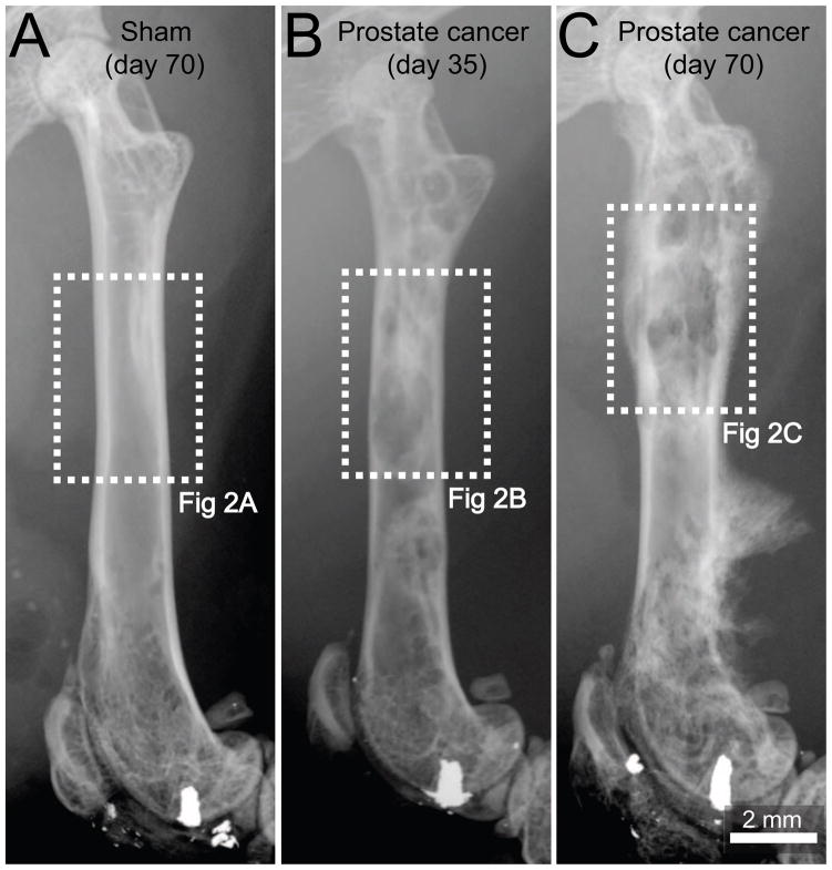
Figure 1. Pathological bone remodeling induced by prostate cancer cells growing within the femur.
Representative radiographic images of a mouse femur at day 70 following injection of culture medium alone (sham, A), at day 35 following injection of ACE-1 prostate cancer cells (B) or day 70 following injection of prostate cancer cells (C). At day 35 post-cancer cell injection the tumor-injected mouse femur shows osteoblastic lesions, characterized by pathological bone formation in the intramedullary space, which generate diaphyseal bridging structures (B). Note that by day 70, the osteoblastic lesions increase in magnitude and an aggressive periosteal reaction is evidenced as a dense and disorganized appearance of the cortical bone in the distal methaphysis and formation of a Codman’s triangle-like structure in the distal diaphysis (C). A Codman’s triangle develops when a portion of periosteum is lifted off of the cortex by tumor and has been reported to occur in bones from patients with metastatic prostate cancer. Sham-injected femurs show no evidence of bone formation or bone destruction (A). Dashed line rectangles in white represent the areas of the bones illustrated in figure 2.

