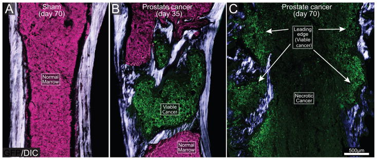
Figure 2. The evolving histopathology as prostate cancer cells grow within the mouse femur.
Differential interphase contrast (DIC) images overlaid on confocal green fluorescent protein (GFP) images (20 μm-thick) of a sham (A) and prostate cancer cell-injected femur from mice sacrificed at days 35 (B) and 70 (C) post-cancer cell injection. DIC images were acquired to visualize cortical and pathological woven bone. As the GFP+ tumor cells (green) grow in normal bone marrow (normal tightly packed hematopoietic cells, pink) they induce the formation of woven bone around the tumor cell colony and eventually form an osteoblastic lesion (B). As tumor disease progresses (day 70), the parent cancer cell colonies undergo necrosis, whereas viable, daughter cancer cell colonies, which are also surrounded by new bone, form at sites more distant from bone marrow (C). Note that at late stages of the disease, the diameter of the femur is greater than that of the femur at day 35 post-tumor injection, or sham bones due to the continuous bone growth induced by prostate cancer cells. Note that osteoblastic lesions are not present in the sham femur (A). For illustration purposes, the background signal which corresponds to healthy bone marrow is presented as the color pink and the GFP+ tumor cells as green.

