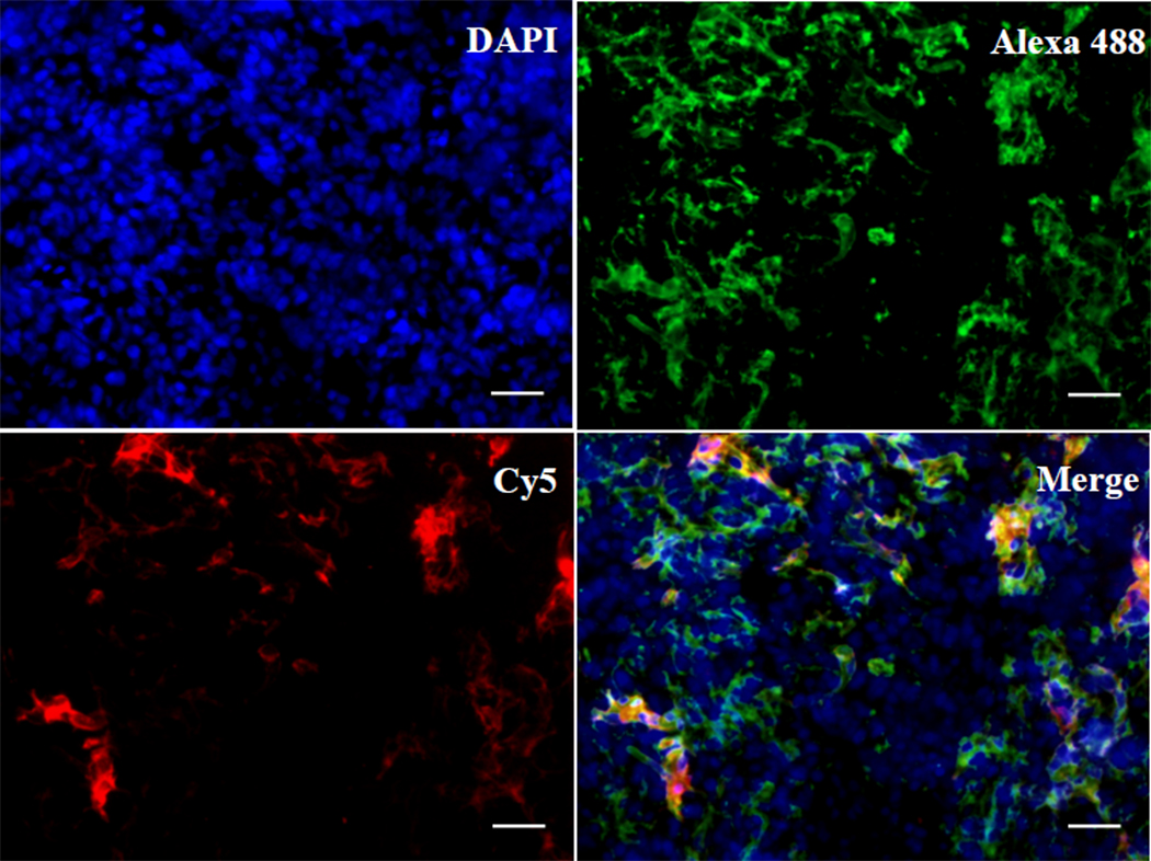
Fig. 2.
Histological analysis of binding of CB-Luc to collagens in tumor. The Luc8-535 and collagen binding protein CNA35 fusion protein CB-Luc was labeled with Cy5 (excitation: 650 nm; emission: 670 nm). The tumor tissues were collected 24 h after i. v. injection of 0.5 nmol of Cy5-labeled CB-Luc. The frozen tissue sections were stained with primary collagen type I antibody and Alexa 488 conjugated secondary antibody (Alexa 488: excitation: 499 nm; emission 519 nm). The Cy5-labeled CB-Luc (red) co-localizes well with type I collagen (green), indicating the accumulation of CB-Luc on the type I collagen after 24 h. Scale bar: 50 µm.

