Fig. 3.
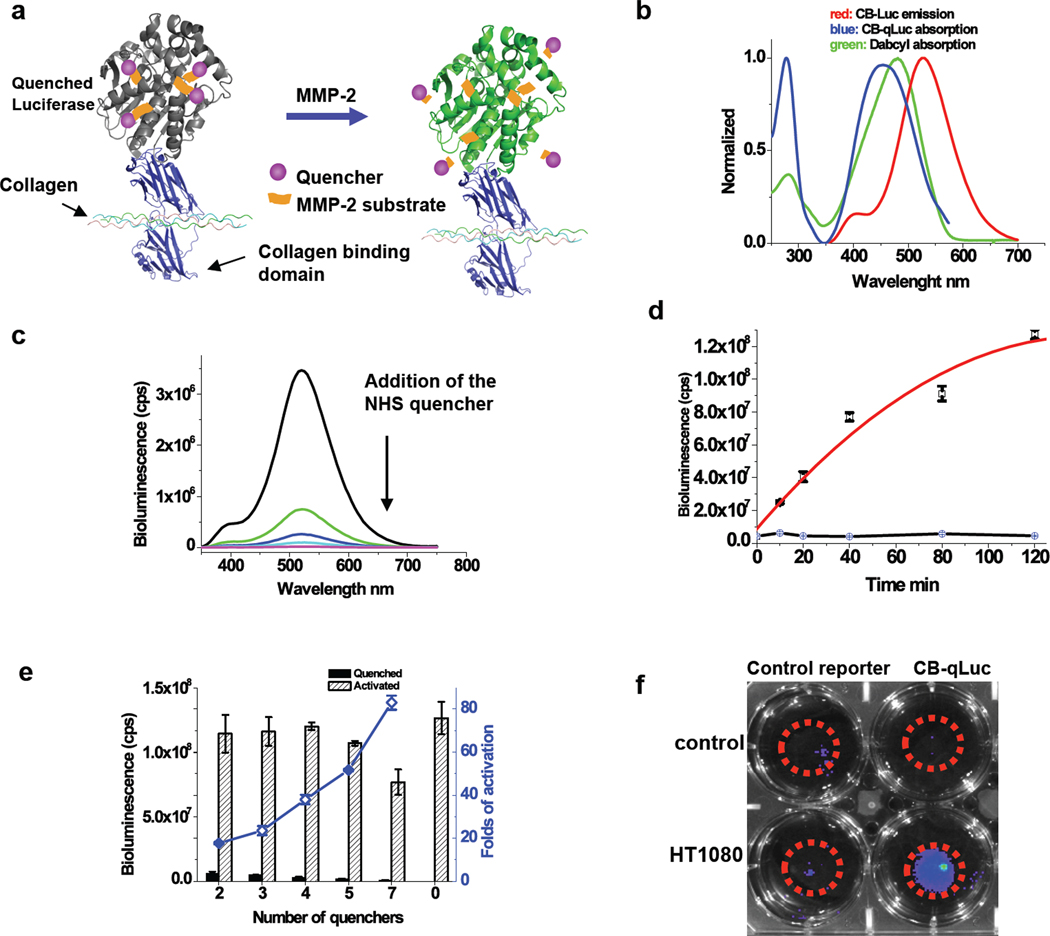
Characterization of bioluminescent activatable reporter CB-qLuc for mapping MMP-2/9 activity in living mice. (a) Design of bioluminescent reporter CB-qLuc for sensing MMP-2/9 activity. (b) Emission spectrum of CB-Luc, and absorption spectra of dabcyl and dabcyl-conjugated CB-qLuc. (c) Bioluminescence emission spectrum of CB-Luc (8 µM in 200 µl PBS buffer, pH 7.4) after a stepwise addition of the NHS ester of dabcyl-conjugated MMP-2/9 substrate in every 15 mins over the course of 1 h. The amount of the quencher in the solution increases from the top to bottom: 0, 2 equivalent, 4 eq., 6 eq. 8 eq.; typically 4 quenchers are conjugated to CB-Luc at the end of the reaction. (d) Activation kinetics of quenched CB-qLuc (red) and the scrambled control reporter (black) (1.0 µM; 4 quenchers per protein on average) by recombinant MMP-2 (160 nM) in 0.05% Tween-20 PBS buffer (pH 7.4) at 37 °C. (e) Effect of the number of quenchers on the luciferase activity of CB-qLuc before and after MMP-2 activation for 2 h under the same condition as in (d). (f) Imaging MMP-2/9 activity in cell culture with 10 pmol of CB-qLuc (right row) or scrambled probe (left row) immobilized in 50 µl matrigel at the center of the dishes (marked by red circles). HT1080 cells (50% confluence) were cultured for 12 h before bioluminescence imaging and no cells were in the control dishes.

