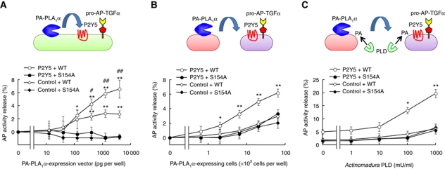
Figure 7.
PA-PLA1α induces P2Y5-dependent TGFα release. PA-PLA1α-mediated activation of P2Y5 in autocrine (A) and paracrine (B, C) manners. (A) Spontaneous AP-TGFα release from HEK293 cells expressing catalytically active PA-PLA1α (WT), P2Y5 and AP-TGFα. Note that AP-TGFα release was not observed in cells expressing catalytically inactive PA-PLA1α mutant harbouring a serine-to-alanine substitution at residue 154 (S154A). Also note that AP-TGFα release was significantly reduced when P2Y5 vector was replaced with an empty vector (control) (n=4; *P<0.05, **P<0.01 versus control vector-transfected cells; #P<0.05, ##P<0.01 versus S154A-expressing cells). (B) PA-PLA1α-induced paracrine activation of P2Y5. P2Y5- and AP-TGFα-expressing cells were mixed with PA-PLA1α-expressing cells that were separately prepared (n=4; *P<0.05, **P<0.01 versus control vector-transfected cells and S154A-expressing cells). (C) Exogenous phospholipase D (PLD) potently enhanced the PA-PLA1α-mediated AP-TGFα release. Same as B (2 × 10³ PA-PLA1α-expressing cells per well), but in the presence of bacterial PLD. AP activity in the absence of PA-PLA1α-expressing cells and PLD treatment was used as the baseline (n=4; *P<0.05, **P<0.01 versus control vector-transfected cells and S154A-expressing cells).

