Figure 1.
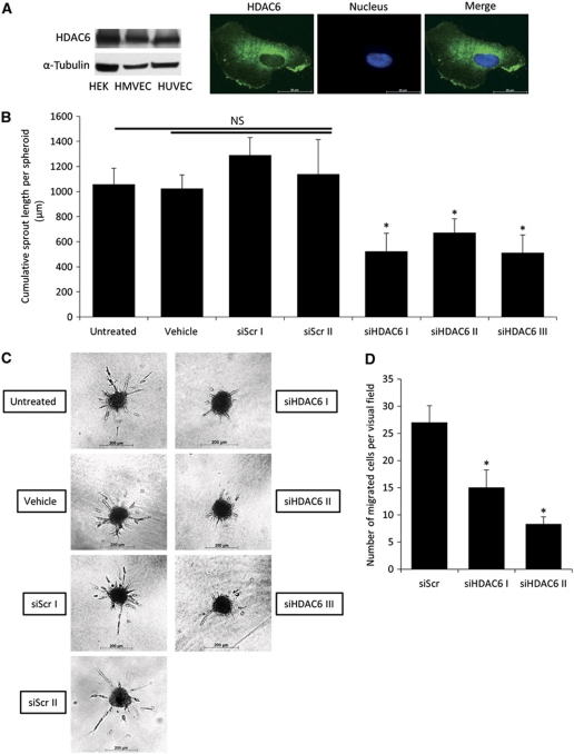
Knockdown of HDAC6 decreases endothelial cell sprouting and migration in vitro. (A) Western blot analysis of HDAC6 expression in cultured endothelial and non-endothelial cells (left panel). α-Tubulin serves as loading control. Immunofluorescence analysis of HDAC6 (shown in green) localization in HUVECs (right panel). Nuclei are stained with Hoechst 33342 in blue. (B) Capillary-like sprouting from spheroids was measured after HDAC6 silencing with three independent HDAC6 siRNAs compared to two different control siRNAs, untreated and vehicle (only transfection reagent)-treated cells. The spheroid assay was performed 24 h after siRNA transfection. Data are shown as mean cumulative sprout length per spheroid (*P<0.05 versus Scr I and Scr II, n=3–10). (C) Representative pictures are shown. (D) HUVECs were transfected with HDAC6 or control siRNA for 48 h, and cell migration was monitored in a transwell migration assay for 4 h under basal conditions. Migrated cells were counted by staining the nuclei with DAPI (n=4). HEK, human embryonic kidney cell.

