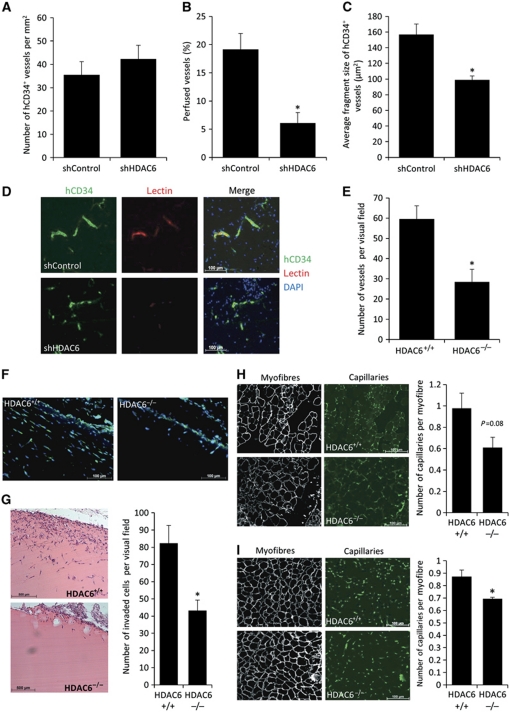
Figure 3.
HDAC6 contributes to human vessel maturation and perfusion in an in vivo mouse model. (A–C) Immunohistological analysis of human neovessel formation after subcutaneous injection of HUVEC spheroids in a matrigel–fibrin matrix in mice. Human vessels were stained with hCD34 antibody. (A) Quantification of microvascular density. (B) Perfusion was analysed as percentage of TRIC-lectin-positive vessels in relation to hCD34-positive vessels. (C) Average fragment size of human vessel was determined measuring the total outline surface of hCD34-positive vessels in relation to the total number of human vessels using ImageJ. (A–C) shControl, n=5; shHDAC6, n=4. *P<0.05. (D) Representative pictures of the human vasculature (shown in green) and perfusion (shown in red). Nuclei are shown in blue. (E) HDAC6 knockout mice (HDAC6−/−, n=6) and wild-type (HDAC6+/+, n=5) mice were subjected to a matrigel plug assay. Lectin-positive structures were counted manually. *P<0.05. (F) Representative images of invaded lectin-positive vessels into the matrigel plug. (G) Quantification of invaded cells into the matrigel plug by H&E staining (right panel, n=5 each). Left panel shows representative images. *P<0.05. (H, I) HDAC6 knockout mice (HDAC6−/−) and wild-type (HDAC6+/+) mice were subjected to hind limb ischaemia. Capillary density was examined histologically in the thigh (H; n=4 for HDAC6+/+ versus n=6 for HDAC6−/−) and the lower leg (I; n=5 each). Left panels show representative images of the capillaries stained for lectin and myofibres stained for laminin. Right panels show the quantification of number of capillaries per myofibre. *P<0.05.

