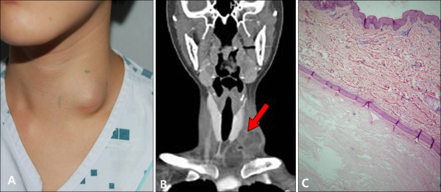
Fig. 2.
(A) A 25-year-old woman with a dome-shaped subcutaneous tumor located on the left lower neck. (B) Computed tomographic scan showed a multilocular cystic mass measuring 6×5×3 cm (arrow). The mass showed expansion toward the trachea. (C) The mass showed a keratinized squamous epithelial lining with the inner surface lined with keratin lamellas (H&E, ×40).

