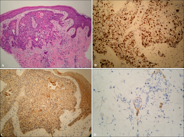
Fig. 2.
Infiltrating tumor nests were observed in the dermis, which were composed of moderately differentiated squamous cells (A: H&E stain, ×100). Tumor cells were positive at Ki-67 (B: ×200) with a high labeling index (≥50%) and pan-cytokeratin staining (C: ×200) with a cytoplasmic pattern; some tumor cells were observed in the lymphatic channel (D: D2-40 stain, ×400).

