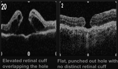
Figure 5.
The optical coherence tomography (OCT) image on the left shows an elevated cuff of subretinal fluid at the margin of the macular hole. According to Hillenkamp et al, it was found to be a strong prognostic indicator for both anatomical closure (p=0.001), and best corrected visual acuity improvement (p=0.048). This is in comparison to the OCT image on the right of a flat, punched out macular hole with no distinct retinal cuff. Reproduced with permission from Hillenkamp J, et al.15

