FIGURE 4.
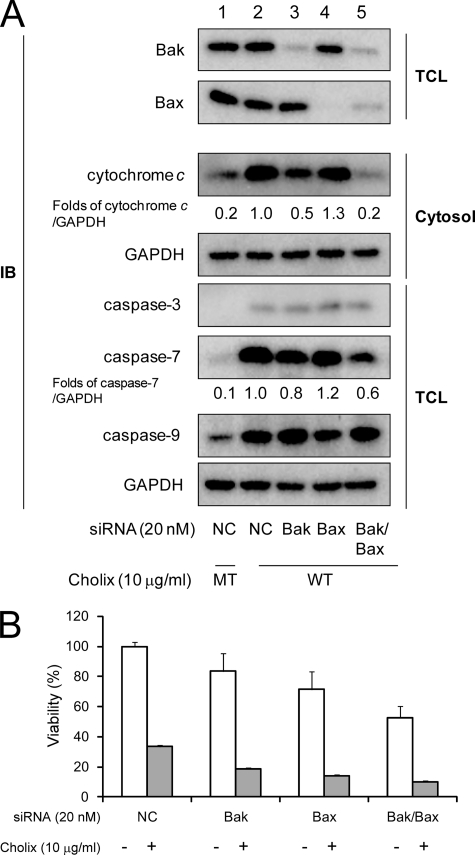
Effect of Bak and/or Bax knockdown on Cholix-induced apoptotic signals. A, cells (1 × 10⁵ cells/well) in a 12-well dish were grown for 24 h, and silencing of bak alone, bax alone, or bak/bax gene was performed with non-targeting control (NC), Bak, or Bax siRNA as described under “Experimental Procedures.” 48 h after transfection, cells were incubated with wild-type (WT) or mutant (MT) Cholix (10 μg/ml) for 18 h. Reduction of Bak or Bax protein level in total cell lysate (TCL) was confirmed by Western blotting with anti-Bak or anti-Bax antibody. Cytochrome c release was determined as described under “Experimental Procedures.” Cholix-induced caspases cleavage was determined by Western blotting with anti-cleaved caspase-3, -7, or -9 antibodies. Mean values were calculated on relative band intensity in three separate experiments. B, the cells transfected with the siRNA overnight (1 × 10⁴ cells/well) were plated in a 96-well dish, grown overnight, and then treated with Cholix (10 μg/ml) for 24 h. Cell viability was determined by using the Cell Counting kit as described under “Experimental Procedures.” Data are the means ± S.D. value from three separate experiments. The Student's t test was used for comparisons with the non-targeting control siRNA-transfected samples incubated with Cholix.

