FIGURE 5.
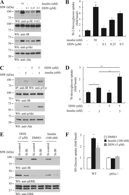
DDN enhances cellular glucose uptake. A, DDN provokes IR signaling in differentiated 3T3-L1 adipocytes. The 3T3-L1 cells were stimulated with insulin (50 nm) for 15 min or DDN (0.1, 0.25, and 0.5 μm) for 30 min. Insulin signaling in the cell lysates was tested by immunoblotting. B, DDN stimulates glucose uptake. Differentiated 3T3-L1 adipocytes were stimulated with insulin (50 nm) for 15 min or DDN (0.1, 0.25, and 0.5 μm) for 30 min. [2-3H]Deoxyglucose was then added, and the cells were further incubated for 10 min. [2-3H]Deoxyglucose uptake by adipocytes was measured by scintillation counting (*, p < 0.05; **, p < 0.01 versus control, Student's t test, n = 3). C, DDN synergizes insulin signaling. Differentiated 3T3-L1 adipocytes were stimulated with 5 nm insulin, 5 nm DDN, or a combination of the two drugs. Cell lysates were then prepared for Western blot using specific antibodies as indicated. D, DDN synergizes insulin activity in promoting glucose uptake. Differentiated 3T3-L1 adipocytes were stimulated with 5 nm insulin, 5 nm DDN, or a combination of the two drugs. [2-3H]Deoxyglucose was then added, and the cells were further incubated for 10 min. [2-3H]Deoxyglucose uptake by adipocytes was measured by scintillation counting (*, p < 0.05; **, p < 0.01, Student's t test, n = 3). E, IR is necessary for DDN to induce ERK phosphorylation. Differentiated 3T3-L1 adipocytes were transfected with control siRNA or siRNA against IR. The cells were then stimulated with insulin (100 nm) for 15 min or DDN (5 μm) for 30 min. Cell lysates were analyzed by Western blot. F, the glucose uptake induced by DDN in p85α knock-out MEF cells. Wild-type and p85α−/− MEF cells were treated with 100 nm insulin for 15 min or 5 μm DDN for 30 min. The uptake of [2-3H]deoxyglucose was monitored by liquid scintillation counting (*, p < 0.05; **, p < 0.01 versus control, Student's t test, n = 3).

