FIGURE 4.
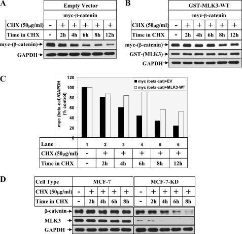
MLK3 regulates β-catenin expression at a post-translational level. HeLa cells transfected with myc-β-catenin in combination with empty vector (A) or GST-tagged MLK3-WT (B) were treated with 50 μg/ml CHX for the indicated periods of time after recovery in growth medium. The first lane represents the samples harvested without any CHX treatment. Western blot analysis was performed with the antibodies indicated. C, the bar graph shows the ratio of myc-β-catenin and GAPDH obtained from the Western blots of A and B considering the respective CHX untreated values as 100%. D, MCF-7 or MCF-7-KD (with stable MLK3 knockdown) were treated with CHX as in A and analyzed by Western blots with the indicated antibodies.

