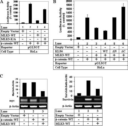
FIGURE 6.
MLK3 inhibits β-catenin and TCF-mediated transcriptional activity. A, HeLa cells were transiently transfected with pGL3OT luciferase (pGL3OT) reporter and β-galactosidase vector (for normalization) along with empty vector (lane 1), β-catenin alone (lane 2), or β-catenin in combination with MLK3-WT (lane 3) or MLK3-K/A (lane 4). Luciferase and β-gal assays were performed as described elsewhere (19) considering the empty vector values as 100%. B, HeLa cells were transfected with pGL3OT and β-gal along with either empty vector (lane 1), β-catenin alone (lane 2), or β-catenin in combination with MLK3-WT (lane 3) or MLK3-WT and KLF4 vectors (lanes 4–6). Luciferase and β-gal assays were performed as in A. Each transfection (A and B) was performed in triplicate, and the data represent the mean ± S.D. of at least two independent experiments. C, shown is a semiquantitative PCR analysis of HeLa cells transfected with empty vector, β-catenin alone, or in combination with MLK3-WT to detect myc or Survivin expression. Actin was used as a control. The bar graphs represent the ratio of myc/β-actin and Survivin/β-actin.

