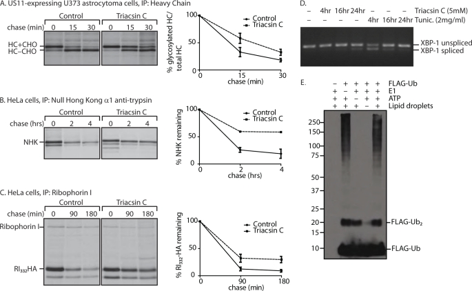
FIGURE 7.
Pharmacological inhibition of lipid droplet formation affects dislocation. A,US11-expressing astrocytoma cells were treated with 5 μm triacsin C for 24 h. Cells were pulse-labeled with 35S-labeled cysteine and methionine. Samples were taken at the indicated chase times, and class I MHC heavy chain was recovered from the lysates. Immunoprecipitates (IP) were separated by SDS-PAGE and imaged by autoradiography. Amount of recovered protein was quantified by phosphorimagery and is shown as a percentage of glycosylated heavy chain compared with total heavy chain. Error bars represent standard deviation of three individual experiments. Solid line, control; dashed line, triacsin C-treated cells. B, HeLa cells were transfected with NHK, and the same experiment as in A was performed, using anti-α1-antitrypsin antibody for immunoprecipitation. C, HeLa cells were transfected with RI332-HA, and the same experiment as in A was performed, using anti-ribophorin I antibody for immunoprecipitation. Quantification in B and C shows the percentage of protein remaining compared with the amount recovered at the 0-min chase time. D, HeLa cells were treated with 5 μm triacsin or 2 μg/ml tunicamycin for the indicated times. RNA was purified and reverse-transcribed. XBP-1 cDNA was amplified by PCR, run on a 2% agarose gel, and visualized by UV. E, lipid droplets were isolated from oleic acid-fed 293T cells by flotation through a sucrose gradient. The lipid droplet fraction was added to an in vitro ubiquitylation assay with 60 μm FLAG-Ub, 100 nm E1, and ATP-regenerating mixture as indicated and incubated for an hour at 37 °C. Lipid droplets were solubilized in a 37 °C sonicating water bath for 2 h, run on a 8% Tris-Tricine gel, and immunoblotted with anti-FLAG antibody.

