FIGURE 3.
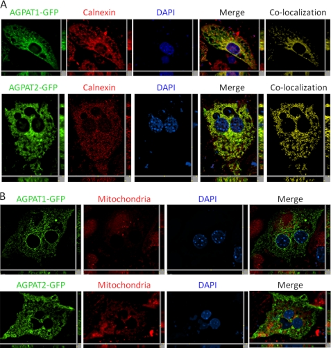
Localization of human AGPAT1-EGFP and AGPAT2-EGFP to endoplasmic reticulum in mouse primary hepatocytes. A, mouse primary hepatocytes with exogenously expressed human AGPAT1-EGFP and human AGPAT2-EGFP were fixed in methanol and incubated with antibody calnexin (specific for endoplasmic reticulum) and imaged for green and red fluorescence using fluorescence microscopy. Shown are representative images for AGPAT1 and AGPAT2 (green fluorescence), calnexin (red fluorescence), DAPI (blue fluorescence), co-localization channel (yellow fluorescence), and the merged image. B, AGPAT1-EGFP- and AGPAT2-EGFP-expressing cells were incubated with MitoTracker Red dye, fixed in 4% paraformaldehyde, and imaged as before. Shown for each image is a single z-stack image, and the x and y axis shows the z-stacks. Scale bar, 5 μm.

