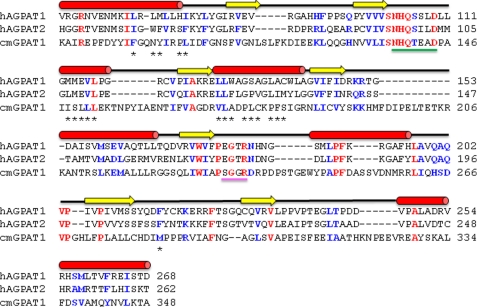
FIGURE 8.
Primary and secondary structure alignment of human AGPAT1 and AGPAT2. Shown are the primary structure alignments of human AGPAT1 (NP_006402.1) and AGPAT2 (CAH71722.1) and squash GPAT (C. moscata BAB17755.1). The secondary structure above the sequences corresponds to that of the AGPAT proteins. α-Helices are colored red; β-sheets are in yellow, and black lines represent coils. The amino acids identified by homology modeling in the hydrophobic tunnel are shown with asterisk. Underlined in green is the catalytic site, with the histidine and aspartate highly conserved amino acids. Underlined in magenta is the EGTR conserved region, with the highly conserved glycine also in the GPAT sequence. These two regions and other highly conserved amino acids were used to establish the homology modeling.

