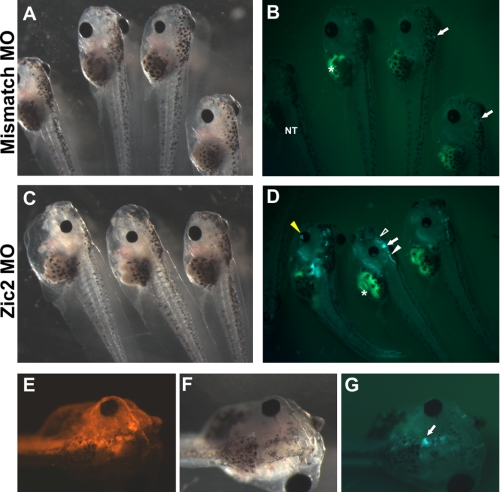
FIGURE 6.
Zic2 morphants display increased Wnt reporter activity in the brain. Lateral views of stage 43 tadpoles injected with 40 ng of mismatch morpholino (MO) show normal development (A) and modest GFP expression (B). Embryos injected with Zic2 morpholino displayed loss of pigmented cells in the head and trunk (C) and markedly increased GFP expression (D), especially in the forebrain (open white arrowhead), the midbrain-hindbrain boundary (arrow), the dorsal side of the hindbrain (closed white arrowhead), and the eyes (closed yellow arrowhead). Note that the intestine shows autofluorescence (asterisk). The left-most tadpole is non-transgenic (NT). E–G, embryo injected unilaterally with Zic2 morpholino together with a red fluorescent LYSAMINE-labeled control morpholino. The injected side shows a marked increase in TOP-GFP expression, especially visible in the midbrain-hindbrain boundary (arrow).

