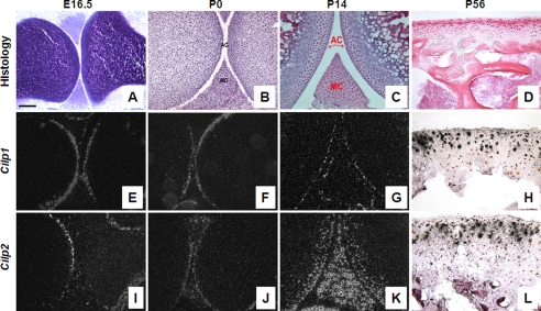
FIGURE 2.
Expression of Cilp1 and Cilp2 mRNA during mouse cartilage development. In situ hybridization on embryonic (E16.5), newborn (P0), 2-week (P14) and 8-week (P56) postnatal mouse femurs. Articular cartilage morphology of mouse femur and tibia stained with toluidine blue (A) or hematoxylin and eosin (B–D) at different time points during development: E16.5 (A), P0 (B), P14 (C), and P56 (D). Dark and bright field images indicate that Cilp1 (E–H) and Cilp2 (I–L) are expressed in the articular and meniscal cartilage. No hybridization with the sense probe was detected (data not shown). AC, articular cartilage; MC, meniscal cartilage. Scale bar, 100 μm.

