FIGURE 6.
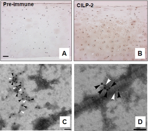
Localization of CILP-2 in human articular cartilage extracts. A and B, in human articular cartilage, CILP-2 protein was detected in the inter-territorial matrix, in the intermediate to deep zone (B). Control sections were probed with CILP-2 preimmune sera (A). Scale bar, 50 μm. C and D, representative micrographs confirming CILP-2 distribution in human articular cartilage visualized by electron microscopy. Immunogold particles (CILP-2, 18-nm particles black arrowheads; collagen VI, 12-nm particles, white arrowheads) were localized within collagen VI containing suprastructures (C), and also as part of collagen VI containing suprastructures in close association with the heterotypic collagen II containing cartilage fibrils (D). Scale bar, 100 nm.

