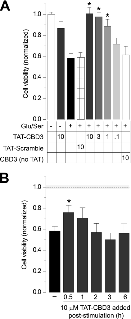
FIGURE 2.
Prevention of glutamate-induced reduction in cell viability by TAT-CBD3. A, cell viability of E18–19 DIV 7 cortical neurons was determined using an MTS cell viability assay. Neurons were incubated with peptides for 10 min prior to stimulation with 200 μm glutamate and 100 μm d-serine for 30 min. Viability was then measured 24 h later, with all values normalized to no stimulation control (n ≥ 32 each from at least four separate experiments). *, significant difference compared with stimulated control (p < 0.05). All values are in μm. B, viability of stimulated neurons was also measured with the addition of TAT-CBD3 at various time points following stimulation (n ≥ 16). *, S.D. compared with stimulated control (p < 0.05). The dotted line and shaded area around the line illustrate the normalized cell viability ± S.E. in the absence of any stimulation (from A). Error bars, S.E.

