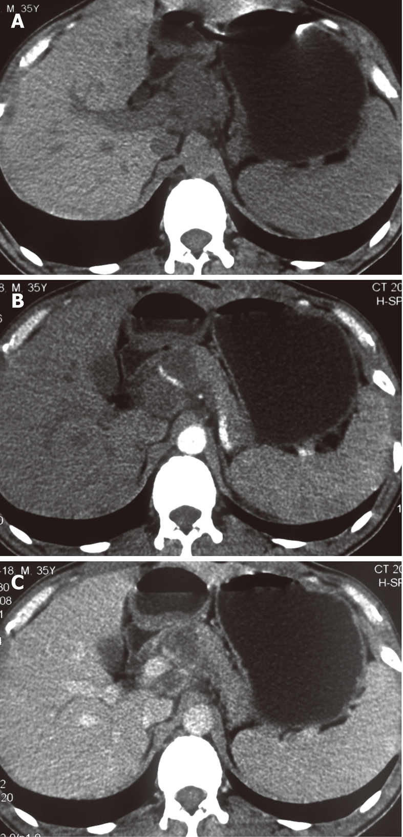
Figure 2.

Peripancreatic tuberculous lymphadenopathy. A: Plain computed tomography (CT) showing peripancreatic, lobular, and low density mass; B, C: Contrast-enhanced CT of the arterial phase (B) and portal venous phase (C) showing the mass with slight peripheral enhancement and the encased common hepatic artery.
