

Abstract
Malignant fibrous histiocytoma (MFH) is the most common soft tissue sarcoma of adults, mostly distributed in the thigh, buttock and groin (46%) and presents rarely in the gastrointestinal tract, classified as gastrointestinal stromal tumors (GIST). MFH is regarded as a diagnosis of exclusion, essentially synonymous with an undifferentiated pleomorphic sarcoma. The recent report presents an 80-year-old man with clinical manifestations of rectal bleeding and final diagnosis of MFH of rectum. It seems that radical resection and adjuvant radiation is important in the management strategy of a patient with such a rare rectal tumor.
Keywords: Malignant fibrous histiocytoma, Gastrointestinal tract, Rectum
1. Introduction
Sarcomas are malignant tumors that arise from mesenchymal tissue at any of the body sites.1 Malignant fibrous histiocytoma (MFH) which was categorized in the 1970s is a malignant pleomorphic stromal tumor for which a definable line of differentiation has not been found. The predisposing factors are genetics, exposure to radiation or chemotherapy, chemical carcinogens, chronic irritation and lymphedema. In nearly all instances, sarcomas are thought to arise de novo and not from a preexisting benign lesion.2,3 The growth pattern of sarcomas is direct local extension, infiltrating adjacent tissues and structures, occasionally with skip areas. Spread to regional nodes is infrequent and vary according to the tumor grade and size.4 Distant metastases often develop later in the course of the disease and are mostly compatible with the histological grade, type and size of the tumor.5 The lungs are ultimately involved in every patient who develops metastatic disease and this represents the predominant cause of death.6
The most common used staging system for soft tissue sarcomas is American Joint Committee on Cancer (AJCC), which is not in widespread use for non-extremity sarcomas.7 Histological grading is another indicator of the degree of malignancy, the probability of distant metastases and death from sarcoma but is a poor indicator of local recurrence. The preferred grading system in these cases is a three level one, including grade I (well differentiated), grade II (moderately differentiated), and grade III (poorly differentiated).1,8 The size and depth of invasion are the most important prognostic factors.9–11
The most common distribution site of MFH is extremities.12 The recent case report is a rare presentation of MFH in rectum.
2. Report of a case
An 80-year-old male was referred to the Department of Surgery, Hazrat-e-Rasoul Hospital, with a 4-month history of rectal bleeding associated with symptoms of fecal incontinency and a loss of weight of about 10 kg. The patient also complained of intermittent bowel obstruction symptoms.
On physical examination the patient was an ill, malnourished man presenting with low rest and squeezing pressure of anal canal. A huge polypoid mass was palpated on the anterior wall of rectum in a distance of six cm to anal verge.
Laboratory tests revealed a moderate anemia (hemoglobin; 9/5 mg/fl), an elevated estimated sedimentation rate (ESR) of 45 mm/h and a serum carcinoembryogenic antigen (CEA) level of 1.9 ng/ml (normal values up to 2.5 ng/ml).
On radiologic investigation, the abdominopelvic CT scan yielded the thickening of the anterior rectal wall without any invasion to adjacent tissues and structures (see Figs. 1 and 2).
Fig. 1.
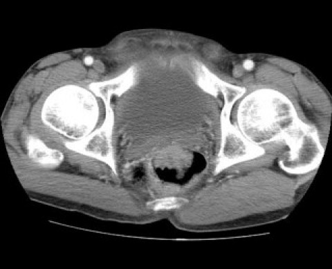
The horizontal cut of the patient's pelvic CT scan.
Fig. 2.
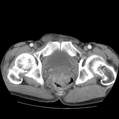
The horizontal cut of the patient's pelvic CT scan.
A biopsy was obtained from the patient in another center which reported a poorly differentiated adenocarcinoma.
The patient's poor general condition, bowel obstruction and rectal bleeding excluded the neoadjuvant chemoradiation therapy as the first line of treatment. The functional impairment of sphincter complex conducted us to plan for an abdominoperineal resection, perineal colostomy and appendicostomy under general anesthesia. The size of the tumor was 5 × 4 × 2.5 cm, 2 cm away from the dentate line.
The histopathological report revealed a highly pleomorphic neoplasm composed of large, pleomorphic giant cells as well as spindle cells, arranged in fascicular and storiform pattern mixed with inflammatory cells infiltration, including lymphocytes, plasma cells, PMNs and multinucleated giant cells (Fig. 3). Immunohistochemical (IHC) staining showed positive reaction of tumor cells with α1 antitrypsin, CD68, Vimentin and focal SMA. Cytokeratin, EMA, CD117(c-Kit) and CD34 were all negative. These findings, in combination with the histomorphology and IHC evaluations, were consistent with malignant fibrous histiocytoma of rectum.
Fig. 3.
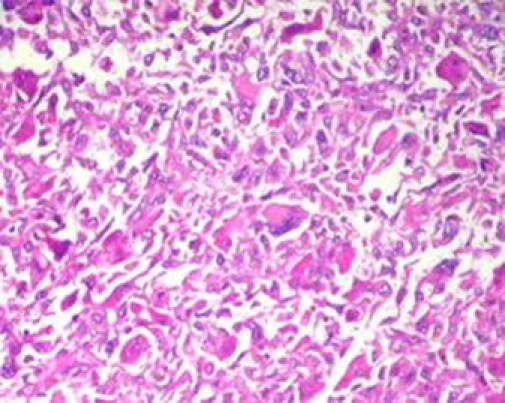
H&E section shows a pleomorphic tumor with sarcomatous features.
3. Discussion
MFH is extremely rare in the gastrointestinal tract, especially rectum. Reviewing the available literatures, we found two cases of MFH of small intestine that were reported by Fu et al. in 1970. The first case was a 70-year-old man with recurrent right lower quadrant pain and a tumor of cecum which was diagnosed as MFH and the second patient was a 43-year-old man who was admitted with intussusception of the small intestine. An emergent laparotomy revealed four pedunculated masses of small intestine which were diagnoses as MFH in the pathology report.13
Waxman et al. reported another case of MFH in colon on 1983, in which the neoplasm was considered as an incidental finding during the diagnostic workup for diverticulitis of the sigmoid colon.14
We found no more than one case of MFH of rectum reported by Dr. Singh et al. from India.15 The patient was a 55-year-old male presented with tennesmus and significant weight loss, who underwent abdominoperineal resection and adjuvant chemoradiation therapy.
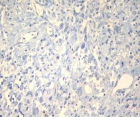
The most probable differential diagnosis of the recent case was Gastrointestinal Stromal Tumor (GIST) which was ruled out by negative staining with c-Kit (Fig. 4). Negative results for CK (Fig. 5), EMA and Vimentin positivity also ruled out the poorly differentiated carcinoma which is the most common malignant neoplasm in this region and sarcomatoid carcinoma. Furthermore diffuse and strong staining with α1 antitripsin (Fig. 6) as well as positive reaction with CD68 (Fig. 7) confirmed the diagnosis of MFH.
Fig. 4.
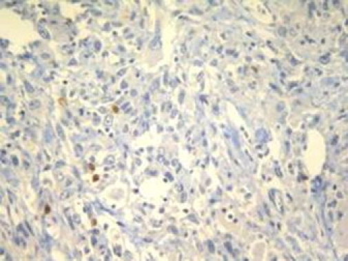
CD117 (c-Kit) is negative in tumor cells.
Fig. 5.
Cytokeratin does not stain tumoral cells.
Fig. 6.
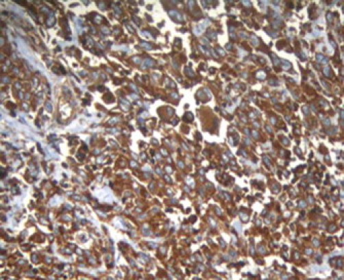
Diffuse and intense positivity of tumor cells with A1-antitrypsin.
Fig. 7.
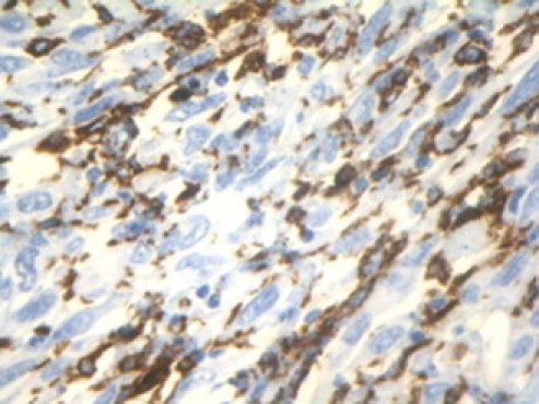
Positivity of some tumor cells with CD68.
Surgical resection is the most potentially effective therapy for soft tissue sarcomas regardless of their site of origin. Adjuvant radiation therapy is recommended for patients with high grade sarcomas beneficial for local control. At present the role of adjuvant chemotherapy for sarcomas remains unclear.3
Conflicts of interest statement
None.
Funding
None.
Patient consent
Written informed consent was obtained from the patient for publication of this case report and accompanying images. A copy of the written consent is available for review
References
- 1.Fletcher C., Unni K., Mertens F. World Health Organization; 2002. Pathology and genetics of tumours of soft tissue and bone. [Google Scholar]
- 2.Raney R.B., Jr., Allen A., O’Neill J., Handler S.D., Uri A., Littman P. Malignant fibrous histiocytoma of soft tissue in childhood. Cancer. 1986;57(June (11)):2198–2201. doi: 10.1002/1097-0142(19860601)57:11<2198::aid-cncr2820571120>3.0.co;2-x. [DOI] [PubMed] [Google Scholar]
- 3.Delaney TF. Overview of soft tissue sarcoma: up to date; 2010.
- 4.Mazeron J.J., Suit H.D. Lymph nodes as sites of metastases from sarcomas of soft tissue. Cancer. 1987;60(October (8)):1800–1808. doi: 10.1002/1097-0142(19871015)60:8<1800::aid-cncr2820600822>3.0.co;2-n. [DOI] [PubMed] [Google Scholar]
- 5.Rydholm A., Berg N.O., Gullberg B., Thorngren K.G., Persson B.M. Epidemiology of soft-tissue sarcoma in the locomotor system. A retrospective population-based study of the inter-relationships between clinical and morphologic variables. Acta Pathol Microbiol Immunol Scand A. 1984;92(September (5)):363–374. [PubMed] [Google Scholar]
- 6.Christie-Large M., James S.L., Tiessen L., Davies A.M., Grimer R.J. Imaging strategy for detecting lung metastases at presentation in patients with soft tissue sarcomas. Eur J Cancer. 2008;44(September (13)):1841–1845. doi: 10.1016/j.ejca.2008.06.004. [DOI] [PubMed] [Google Scholar]
- 7.Greene F. Springer Verlag; 2002. AJCC cancer staging manual. [Google Scholar]
- 8.Deyrup A.T., Weiss S.W. Grading of soft tissue sarcomas: the challenge of providing precise information in an imprecise world. Histopathology. 2006;48(January (1)):42–50. doi: 10.1111/j.1365-2559.2005.02288.x. [DOI] [PubMed] [Google Scholar]
- 9.Rooser B., Willen H., Gustafson P., Alvegard T.A., Rydholm A. Malignant fibrous histiocytoma of soft tissue. A population-based epidemiologic and prognostic study of 137 patients. Cancer. 1991;67(January (2)):499–505. doi: 10.1002/1097-0142(19910115)67:2<499::aid-cncr2820670230>3.0.co;2-e. [DOI] [PubMed] [Google Scholar]
- 10.Rydholm A., Syk I. Malignant fibrous histiocytoma of soft tissue. Correlation between clinical variables and histologic malignancy grade. Cancer. 1986;57(June (12)):2323–2324. doi: 10.1002/1097-0142(19860615)57:12<2323::aid-cncr2820571214>3.0.co;2-r. [DOI] [PubMed] [Google Scholar]
- 11.Weiss S.W. Malignant fibrous histiocytoma. A reaffirmation. Am J Surg Pathol. 1982;6(December (8)):773–784. doi: 10.1097/00000478-198212000-00009. [DOI] [PubMed] [Google Scholar]
- 12.Lawrence W., Jr., Donegan W.L., Natarajan N., Mettlin C., Beart R., Winchester D. Adult soft tissue sarcomas. A pattern of care survey of the American College of Surgeons. Ann Surg. 1987;205(April (4)):349–359. doi: 10.1097/00000658-198704000-00003. [DOI] [PMC free article] [PubMed] [Google Scholar]
- 13.Fu D.L., Yang F., Maskay A., Long J., Jin C., Yu X.J. Primary intestinal malignant fibrous histiocytoma: two case reports. World J Gastroenterol. 2007;13(February (8)):1299–1302. doi: 10.3748/wjg.v13.i8.1299. [DOI] [PMC free article] [PubMed] [Google Scholar]
- 14.Waxman M., Faegenburg D., Waxman J.S., Janelli D.E. Malignant fibrous histiocytoma of the colon associated with diverticulitis. Dis Colon Rectum. 1983;26(May (5)):339–343. doi: 10.1007/BF02561712. [DOI] [PubMed] [Google Scholar]
- 15.Singh D.R., Aryya N.C., Sahi U.P., Shukla V.K. Malignant fibrous histiocytoma of the rectum. Eur J Surg Oncol. 1999;25(August (4)):447–448. doi: 10.1053/ejso.1999.0677. [DOI] [PubMed] [Google Scholar]