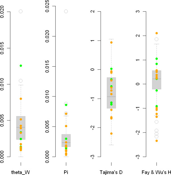
Figure 2.

Box plots summarizing patterns of nucleotide variation in Medicago truncatula. Boxplots (shaded gray) depict the empirical distribution obtained for control fragments. Dots represent individual flowering candidate genes (orange) and symbiotic genes (green). A: Distribution of the scaled mutation rate (as estimated with Watterson's θ) per bp for each fragment. B: Pairwise nucleotide diversity (π). C: Tajima's D statistic for each fragment. Z: standardized Fay and Wu's statistic.
