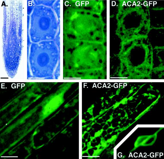
Figure 7.
Imaging of ACA2-GFPp fluorescence reveals ER-like structures in root cells. A, Longitudinal section from a primary root shows undifferentiated cells at the root tip. Cells are stained with toluidine blue to emphasize densely stained nuclei. Toluidine blue-stained sections were photographed on an inverted microscope (model IMT2, Olympus) using Kodak Ektachrome P1600. B, Two undifferentiated cells from A are shown at higher magnification to emphasize the position of the large central nuclei. These cells are representative of live cells imaged for GFP fluorescence in C and D. C and D, Confocal images of green fluorescence from two undifferentiated cells expressing a GFPp-only control (C) or ACA2-GFPp (D). E and F, Green fluorescence from mature root epidermal cells expressing a GFPp-only control (E) or ACA2-GFPp (F), as imaged by computational optical-sectioning microscopy. G, The nuclear region from two neighboring epidermal cells expressing ACA2-GFPp. Imaging of the nuclear region required shorter exposure times, which left the image of the surrounding network only faintly fluorescent. The thickness and angle of this section does not show the cell wall, which lies at an oblique angle between two nuclei in two separate epidermal cells. Scale bars = 50 μm in A; 5 μm in B, C, and D; and 10 μm in E, F, and G. Images were arranged using Adobe PhotoShop (Adobe Systems, Mountain View, CA).

