
Figure 4.
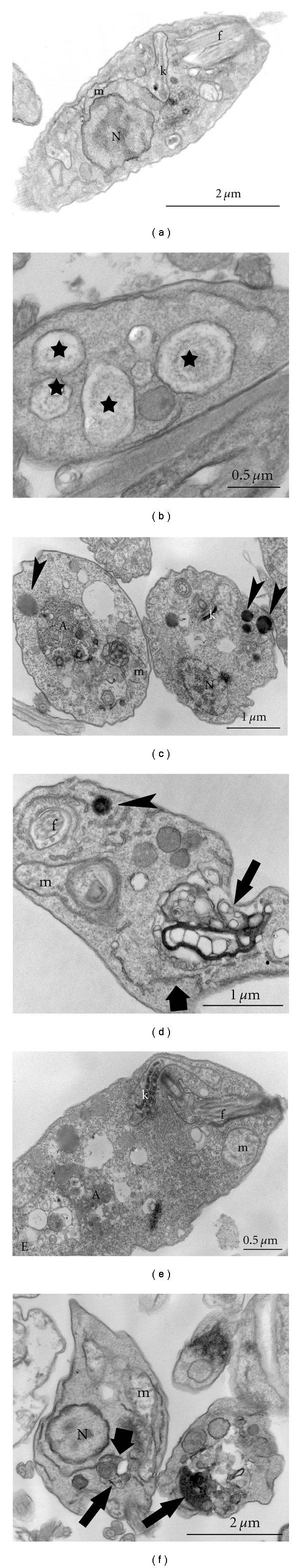
Different ultrastructural alterations on Leishmania amazonensis promastigotes induced by the treatment with amiodarone (AMIO). (a) Ultrathin section of L. amazonensis promastigotes without treatment, which presents a normal ultrastructure of organelles such as (mitochondrion) m, (kinetoplast) k, (nucleus) N and (flagellum) f. (b) Electron micrograph of L. amazonensis treated with 5 μM AMIO for 48 h presenting many vacuoles similar to autophagosomes (stars). (c–e) After treatment with 15 μM AMIO for 24 h, it is possible to observe the presence of large autophagosomes a associated with endoplasmic reticulum profiles (big arrow), lipid bodies (arrowheads), and alterations in the mitochondrion–kinetoplast complex and chromatin condensation. (f) Promastigotes treated with 20 μM AMIO for 24 h presented drastic alterations and destruction of the cytoplasm, where it is possible to observe the presence of autophagosomes (arrows) sometimes associated with endoplasmic reticulum profile (big arrow). A: autophagosome; f: flagellum; k: kinetoplast; m: mitochondrion; N: nucleus.