
Figure 6.
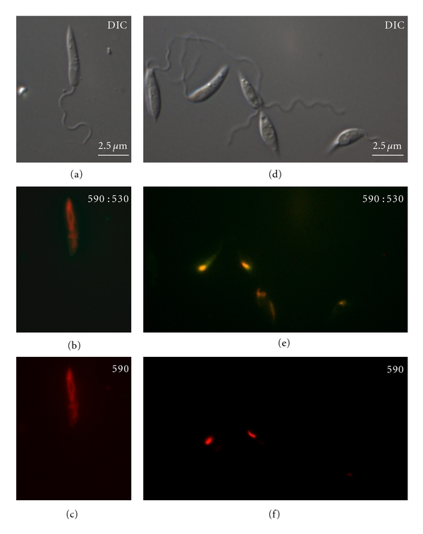
Differential interference contrast (DIC) microscopy and fluorescence microscopy with JC-1 of Leishmania amazonensis promastigotes untreated (a–c) and treated with 15 μM AMIO for 48 h (d–f). In panels (b) and (c), the accumulation of aggregated-JC1 is observed in the whole extension of the control mitochondrion. In cells treated with 15 μM AMIO (e-f), the accumulation of J-aggregates occurs in some portions of the mitochondrion, indicating a partial dissipation of the ΔΨm. Panels (b) and (e) show an image of the monomers and J-aggregates together, while panels (c) and (f) show the image of only J-aggregates.