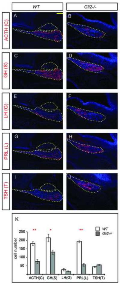
Figure 2.
The number of corticotropes, somatotropes and lactotropes is reduced in Gli2 mutants at E17.5. (A-J) Immunostaining of different pituitary cell type markers ACTH, GH, LH, PRL and TSH at in WT and Gli2 mutant pituitaries. Outlined areas in WT represent anterior and posterior pituitaries. In Gli2 mutants, only anterior pituitary is present. (K) Quantification of each pituitary cell type in WT and Gli2 mutants (* indicates P<0.05, ** indicates P<0.01, n=6 embryos). Two mid-sagittal sections from each embryo were used to quantify the number of different cell type.

