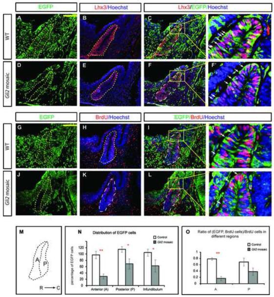
Figure 3.
In mosaics, Gli2 mutant cells do not contribute as well in the anterior edge of the pituitary due to reduced proliferation there. (A-F) Immunostaining of Lhx3 in WT and Gli2 mosaic embryos. (C’,F’,I’,L’) are higher magnification images of corresponding boxes indicated. Note: EGFP is expressed in cytoplasm while Lhx3 and BrdU are present in nuclei. (G-L) Immunostaining of BrdU and EGFP in WT and Gli2 mosaic embryos. EGFP+ cells in WT embryos (A-C, G-I) were Gli2+/+ while EGFP+ cells in Gli2 mosaic (D-F, J-L) were Gli2zfd/lacki. (M) Schematic representation of the subdivisions of the pituitary. R, rostral; C, caudal. (N) Distribution of EGFP+ cells in anterior wall (A) and posterior wall (P) of the anterior pituitary, and in infundibulum. (O) Distribution of BrdU+;EGFP+/BrdU+ cells in different regions of the anterior pituitary (PA<0.01; PP=0.105). See Material and Methods for normalization of the number of EGFP+ cells in each embryo. The anterior pituitary was outlined by dotted lines. White arrowheads indicate cells double-labeled with both green and red. Scale bars: 100 μm. * represents P<0.05, ** represents P<0.01.

