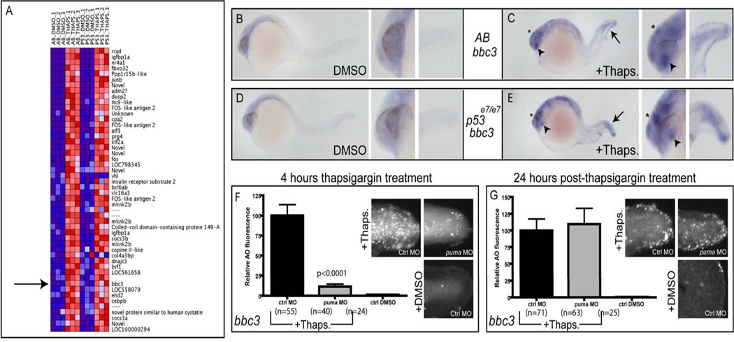
Figure 4. puma expression is increased in a p53-independent manner following thapsigargin treatment.
(A) Microrray analysis of dissected tail tissue revealed increased puma expression following thapsigargin treatment in both AB and p53 mutant embryos. (B,C) Compared to DMSO-treated controls (B), thapsigargin-treated embryos (C) had increased puma expression in the tail (arrow), epiphysis (asterisk), and lens (arrowhead). This increased expression was also observed in thapsigargin-treated p53 mutant embryos (E) compared to DMSO-treated controls (D). (F) Knockdown of puma attenuated 4-hour ER stress-induced apoptosis, but not (G) 28 hour ER stress-induced apoptosis (See also Supplemental Figures 2 and 3).

