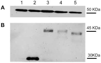
Figure 3. Western Blot analyses of constructs used for cell transfection.
The integrity of the constructs used to transfect cells (shown in Figure 2) was assessed by Western Blotting. After separation of the proteins in cell lysates by SDS-PAGE, the bands were transferred to nitrocellulose membranes and for detection with anti-EGFP and anti-tubulin antibodies followed by peroxidase-labeled anti-rabbit IgG. The protein bands were detected by chemiluminescence. 1. Lysed Huh 7 cells; 2-Huh 7 cells transfected with pEGFPN1, 3-Huh7 cells transfected with core sequence aa(1-173) inserted into pEGFPN1; 4-Huh 7 cells transfected with the core aa(1–160) sequence in pEGFPN1; Huh 7 cells transfected with core sequence aa(1–140) inserted into pEGFPN1.

