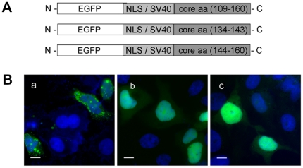
Figure 7. Core protein fragment aa(134–160) contains no other functional NES.
(A) Three plasmids encoding the core fragments aa(109–160), aa(134–143) and aa(144–160) fused to EGFP were constructed as described in the Materials and Methods section. (B) Analysis of the subcellular distribution of the corresponding proteins encoded by the plasmids shown in (A). Cells were transfected with plasmids encoding corresponding EGFP-labeled proteins, grown for 24 h, fixed in 4% PFA and analyzed by fluorescence microcopy. Core protein fragment aa(109–160) was located in the cytoplasm (a), whereas proteins corresponding to core fragments aa(134–143) (b) and aa(144–160) (c) remained in the nucleus. Staining of the nuclei with DAPI. Bar represents 10 µm.

