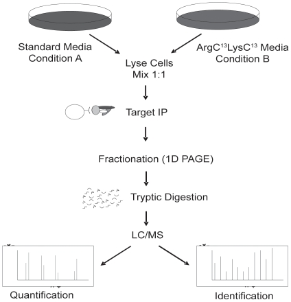
Figure 3. Schematic of stable isotope labeling with amino acid in cell culture and immuno-affinity precipitation for MS analysis (SILAC-IAP-MS).
T293 cells grown in either light (12C-Arg, 12C-Lys) or heavy (13C-Arg, 13C-Lys) medium were lysed and combined at a 1∶1 ratio based on total protein quantity. PDK1 was immunoprecipitated and analyzed by mass spectrometry. MS raw data were quantified by the Elucidator software 3.5 and confirmed by manual validation as described in the Materials and Methods.

