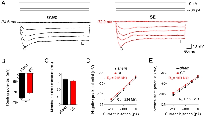
Figure 2. Subthreshold electrophysiological properties of CA1 pyramidal cells.
Individual CA1 pyramidal cells in a hippocampal slice preparation were electrophysiologically characterized in the whole-cell patch-clamp configuration. A. Current-clamp recordings (test pulses of 0 to −200 pA for 900 ms as indicated) obtained from a CA1 pyramidal cell of a sham-treated animal (sham, black traces on the left) and from a CA1 pyramidal cell of a kainate-injected animal with SE behavior (SE, red traces on the right). Note that the potential measured with 0 pA current injection is slightly less negative in the SE cell. Time points for the measurement of negative peak potential (circles) and steady-state potential (squares) are indicated. B. Resting potential of sham (black bar, n = 65) and SE cells (red bar, n = 81); * p<0.05). C. Membrane time constant (obtained with a single-exponential fit to the initial voltage deflection in response to a −50 pA current injection) of sham (black bar, n = 65) and SE cells (red bar, n = 81). D. Negative peak potential (see circles in A) plotted against current injection for sham (black, n = 65) and SE cells (red, n = 81). E. Steady-state potential (see squares in A) plotted against current injection for sham (black, n = 65) and SE cells (red, n = 81); error bars in D and E are smaller than symbols; linear fits to the data yielded slopes from which input resistance (Rin) was determined (numbers are mean values).

