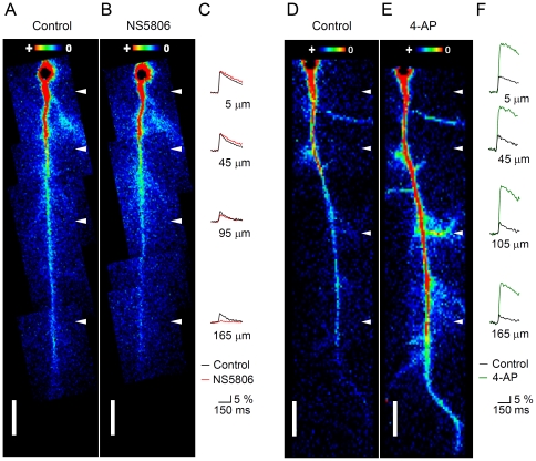
Figure 7. Effect of NS5806 and 4-AP on the dynamics of b-AP-induced Ca2+ signals.
A and B. False color images of a CA1 pyramidal cell from an untreated animal in the absence (Control) and presence of 20 µM NS5806, respectively, during AP backpropagation. The maximal Ca2+ signal (absolute fluorescence change, ΔF) caused by the b-AP is color-coded (insets). C. Corresponding Ca2+ transients (ΔF/F in %) recorded at 5, 45, 95 and 165 µm from the soma (see white arrowheads in A and B) in the absence (black traces) and presence of NS5806 (red traces). Note that the Ca2+ signal at 165 µm from the soma was almost below the detection level in the presence of NS5806. D and E. CA1 pyramidal cell in the absence (Control) and presence of 5 mM 4-AP, respectively, during AP backpropagation. Note that the Ca2+ signal was still visible in the most distal parts of the apical dendrite and invaded lateral branches in the presence of 4-AP. F. Corresponding Ca2+ transients recorded at 5, 45, 105 and 165 µm from the soma (see white arrowheads in D and E) in the absence (black traces) and presence of 4-AP (green traces). Note that the Ca2+ signal showed only little attenuation along the apical dendrite in the presence of 4-AP.

