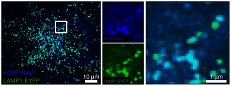
Figure 1. The majority of endo-lysosomal vesicles are positive for both Rab7 and LAMP1.
A representative confocal microscopy image shows the overlaid ECFP-Rab7 (blue) and LAMP1-EYFP (green) images. The inset is split into its individual color components and enlarged. Similar levels of colocalization were observed for BS-C-1 cells stably expressing ECFP-Rab7, for endogenous Rab7 and LAMP1 in BS-C-1 cells, and for HeLa cells (Figure S1).

