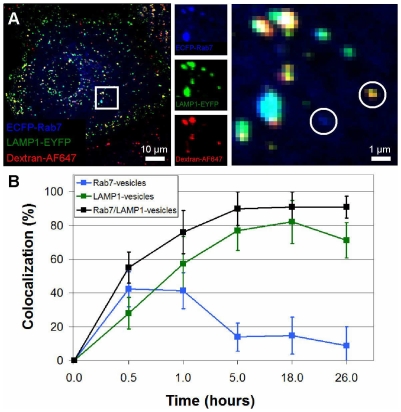
Figure 2. Dextran accumulates in LAMP1- and Rab7/LAMP1-vesicles.
(A) Confocal microscopy image of ECFP-Rab7 (blue), EYFP-LAMP1 (green), and dextran-AF647 (red) after an 18 h incubation with dextran. The inset, split into its three color components and enlarged, shows a Rab7-vesicle in the absence of dextran and a LAMP1-vesicle containing dextran, both circled. (B) At early times dextran is present in Rab7- (blue), LAMP1- (green), and Rab7/LAMP1-vesicles (black). At longer times the percentage of Rab7-vesicles containing dextran decreases while the percentage of LAMP1- and Rab7/LAMP1-vesicles increase. The x-axis is plotted to highlight the early time data. Error bars represent standard deviation. At each time point, analysis was carried out for 10-15 vesicles per cell in 6–12 cells in 2–4 distinct experiments.

