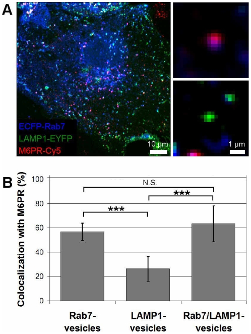
Figure 3. Colocalization of M6PR with Rab7-, LAMP1-, and Rab7/LAMP1-vesicles.
(A) Confocal microscopy image of ECFP-Rab7 (blue), LAMP1-EYFP (green), and an antibody against M6PR labeled with a Cy5-labeled secondary antibody (red). Smaller images show a M6PR-positive (red) Rab7-vesicle (blue, top) and a M6PR-negative LAMP1-vesicle (green, bottom). (B) A large fraction of Rab7- and Rab7/LAMP1-vesicles are positive for M6PR; 57±7% and 63±15%, respectively. A smaller fraction of LAMP1-vesicles are positive for M6PR, 23±10%. Error bars show standard deviations. P-values<0.001 are indicated by ***. N.S. indicates a p-value >0.05. The graph shows the analysis of 10–20 of each type of vesicle per cell in 10 cells from 4 distinct experiments. Similar results were obtained for BS-C-1 cells stably expressing ECFP-Rab7 and for HeLa cells (Figure S4). Unmerged images are shown in Figure S5.

