
Figure 3.
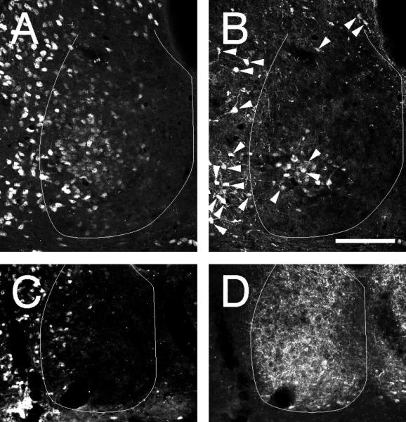
Coronal sections through the mid-caudal hamster SCN showing double label in neurons immunoreactive to(A) NeuN and (B) CalB in the SCNce and for (C) NeuN and (D) VIP-IR. All cells in (B) that show clear co-localization (arrowheads) of CalB- and NeuN-IR. No cells co-localize NeuN- and VIP-IR and the distributions overlap very little. The SCN border line is outlined in white. Bars = 100 μm.