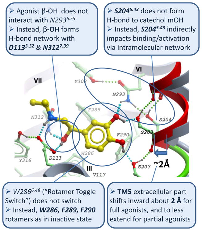
Figure 1.
Key new features of full agonist isoproterenol binding and conformational changes in β2AR ligand binding pocket predicted in 2008 by conformational modeling based on crystal structure of inactive structure of the β2AR-carazolol complex (PDB:2rh1). In this published model (see Supplementary Info in ref. [17]) ligand is shown with yellow carbons, while the contact residues of the β2AR shown with green carbons. Shifted TM5 helix (red and green ribbon) is compared to the original backbone in the crystal structure. Predicted hydrogen bonds are shown by cyan (ligand-receptor) and green (intramolecular) spheres.

