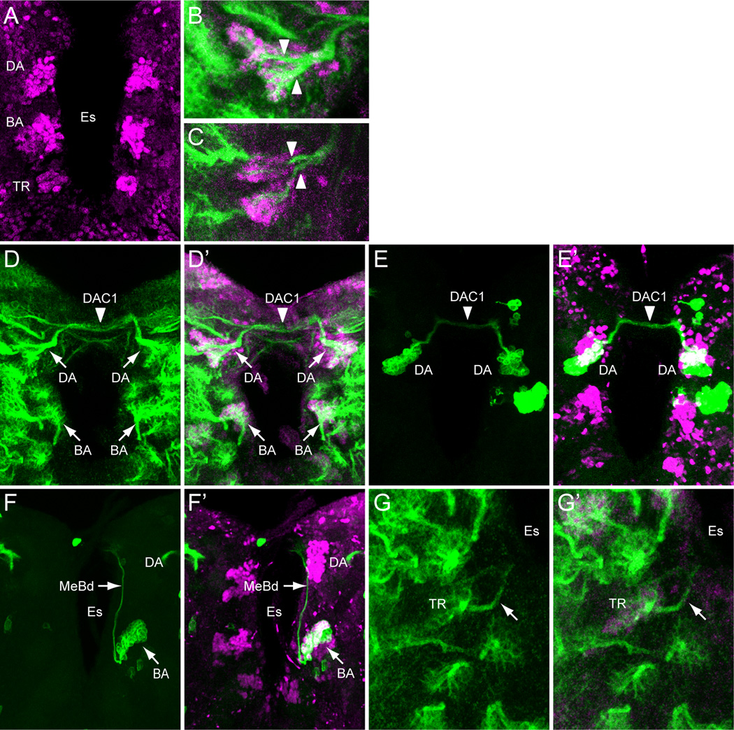
Fig. 2. Identification of sim+ larval brain neurons.
(A) 3rd instar larval brain stained with anti-Sim to show the 3 paired clusters of sim+ neurons: (DA) DAMv1/2, (BA) BAmas1/2, and (TR) TRdm neurons. Esophageal opening (Es) lies between the brain hemispheres. (B,C) Brains stained with anti-Sim (magenta) and MAb BP106 (green) showing (B) DAMv1/2 neurons and (C) BAmas1/2 neurons. Each cluster consists of two groups of neurons that extend an axon (arrowheads) that converge into a single axon tract. (D,D’) Brain stained with anti-Sim (magenta) and MAb BP106 (green) showing neuronal cell bodies and axon tracts. The characteristic tracts of the DAMv1/2 (DA) and BAmas1/2 (BA) neurons are shown (arrows). The DAMv1/2 axons can be seen crossing the midline in the DAC1 axon tract (arrowhead). (E,E’) Brain visualized for GFP (green) and Sim (magenta) showing two DAMv1/2 (DA) MARCM clones that fasciculate together in DAC1 (arrowhead). (F,F’) The characteristic ascending MeBd axon tract (arrow) is shown emanating from a BAmas1/2 (BA) MARCM clone. (G,G’) Brain stained with MAb BP106 (green) and anti-Sim (magenta) illustrating that the sim+ TRdm neurons extend a characteristic short projection (arrow) toward the neuropil near the ventral esophagus (Es).

