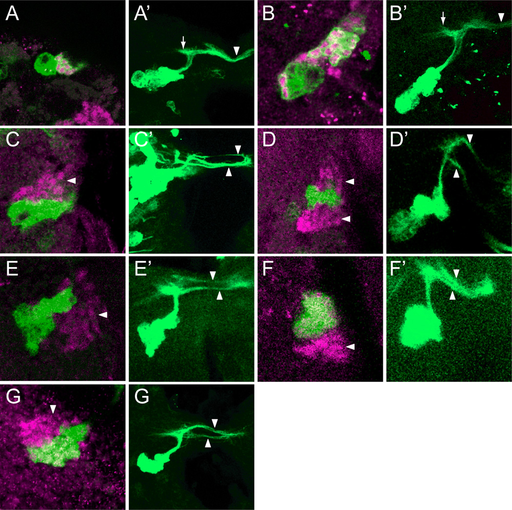
Figure 7. sim mutant clones show axon fasciculation defects.
Wild-type and mutant MARCM GFP+ (green) DMAv1/2 clones were stained with anti-Sim (magenta). For each pair of images, the left panel shows a single optical slice showing the GFP+ cell bodies, and the right panel is a projection that illustrates the axonal morphology. (A,B) Two wild-type GFP+ clones that overlap Sim+ neurons. The NB and GMC are Sim−. (A’,B’) The characteristic DAMv1/2 axon tract is apparent and extends centro-dorsally toward the neuropil, then elaborates filopodia (arrow) before projecting contralaterally (arrowhead) across the supraesophageal commissure. (C,D) Two simBB68 mutant clones that are adjacent to DMAv1/2 Sim+ neurons (arrowheads). (C’,D’) The axons split into multiple fascicles (arrowheads) rather than traverse the supraesophageal commissure as a single, tight fascicle. (E–F’) Two sim2 clones and a (G,G’) sim8 clone that also show multiple branches.

