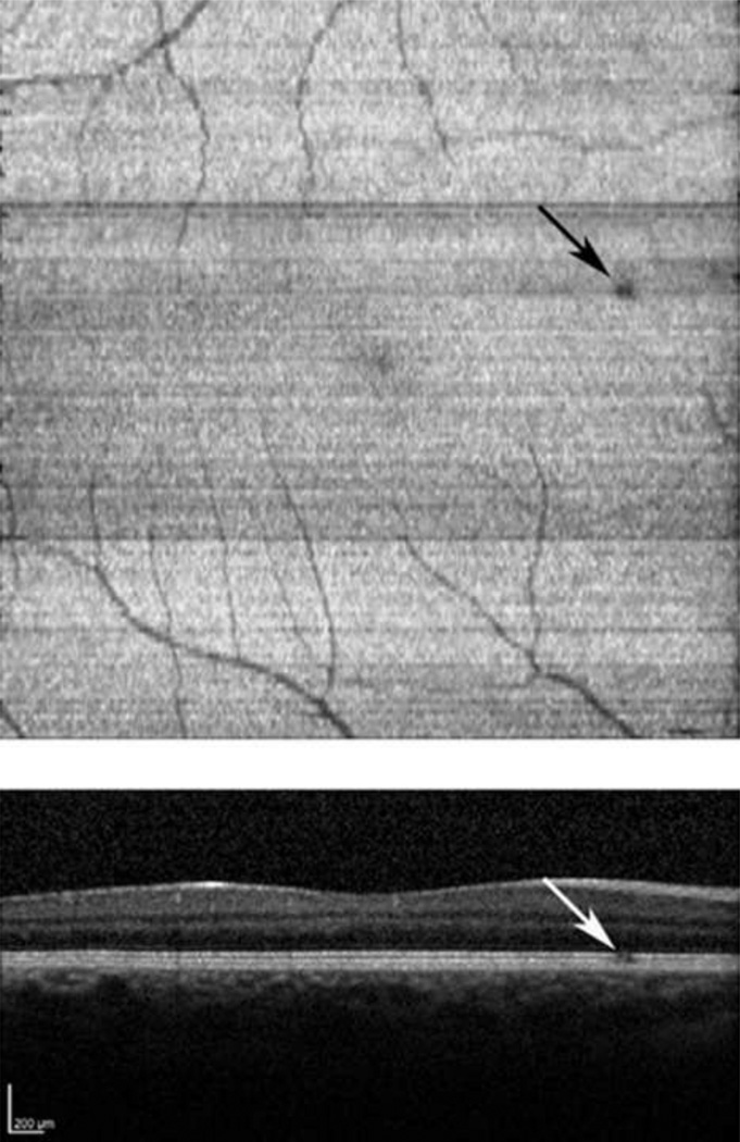
Figure 3.
Normal Subject: (Top) An inner and outer segment enface image from a normal subject, revealing a focal dark region located superonasal to the fovea (black arrow). (Bottom) A SDOCT B-scan traversing the dark region in (Top). A small discontinuity in the inner and outer segment junction is marked with a white arrow.

