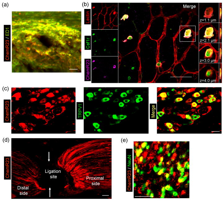
Figure 3.
The RvE1 receptor ChemR23 is widely expressed in immune cells, glial cells, and neurons in mouse tissues. (a) Double staining of ChmR23 and ED1 shows that ChemR23 is largely colocalized with the macrophage marker ED1 in the dermis of the CFA-inflamed skin. Scale bar: 50 μM. (b) Triple staining in retinal whole-mounts demonstrates that ChemR23 is localized to a subset of colony-stimulating factor-1-receptor–positive (Csf1r+) microglia. Left column, triple staining for lectin (endothelial cells and microglia, red), Csf1r (green) and ChemR23 (magenta). Central panel, merged image (scale bar: 50 μm); white indicates the colocalization of all three stains. Right column, four images of one Csf1r+ cell at indicated focal planes. (c) Double staining of ChemR23 and TRPV1 shows that ChemR23 is largely co-localized with TRPV1 in DRG neurons. Note that ChemR23 is also expressed in satellite glial cells surrounding neurons. Scale bar: 30 μM. (d) ChemR23 staining shows that after ligation of the sciatic nerve ChemR23 is accumulated near the ligation site (arrows), indicating axonal transport of ChemR23. Scale bar: 30 μM. (e) Double staining of ChemR23 and NeuN shows that ChemR23 co-colocalizes with the neuronal marker NeuN in the spinal cord dorsal horn. Scale bar: 50 μM. Note that ChemR23 is also expressed in NeuN-negative glial cells in the spinal cord. Reproduced/modified, with permission, from [61] (panel b), [71] (panels a, c–e).

