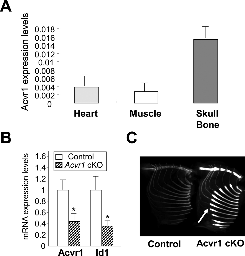
Figure 1.
Acvr1 conditional knockout (cKO) mice. (A) Acvr1 expression levels in normal tissues. RNA was isolated from newborn wildtype mice. Endogenous expression levels of Acvr1 were assessed by qRT-PCR. Absolute values were expressed as mean ± SD (n = 3). (B) qRT-PCR analysis for Acvr1 and Id1 using P21 calvariae. Expression levels of Acvr1 and Id1 were significantly reduced in cKO bones. Values in cKO bones (striped bar) are expressed relative to controls (open bar). mean ± SD; *, p < 0.05. (C) X-ray image of Acvr1 cKO adult mice. The radiodensity of cKO rib bones was notably increased compared with controls. White arrows indicate the rib flaring in cKO mice.

