
Figure 1. Immune reconstitution in a patient with IPEX after non-myeloablative HSCT.
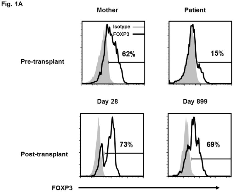
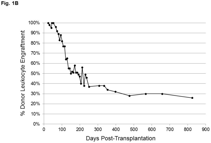
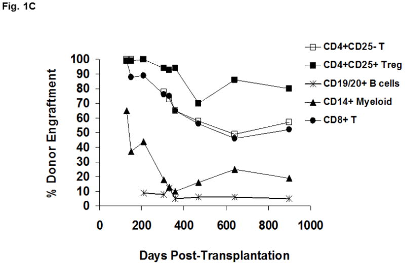
(A) Lymphocytes were selected based on exclusion of monocytes and granulocyes and the CD4+ T cell population was partitioned into the CD4+CD25− and CD4+CD25bright subsets (as shown in Figure 2B and 2C). FOXP3 expression within the CD4+CD25bright T cells (solid line) is compared to an isotype control antibody (shaded area). (B) Donor chimerism of total peripheral blood leukocytes at various times after HSCT, as assessed by VNTR. (C) Multi-color flow cytometry was used to sort lineage-specific cell populations prior to chimerism analysis. Donor chimerism of the purified subsets as determined by VNTR is indicated.