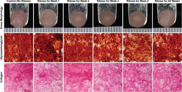
Figure 1.
Gross morphology and histology of self-assembled constructs at 4 weeks. From left to right: representative images of constructs from Control, Week 1, Week 2, Week 3, Week 4, and All Weeks ribose treatment groups. Constructs had a similar flat circular appearance with no surface abnormalities (top row). All constructs stained positively for GAG (middle row) and collagen (bottom row). Color images available in online version of this article.

