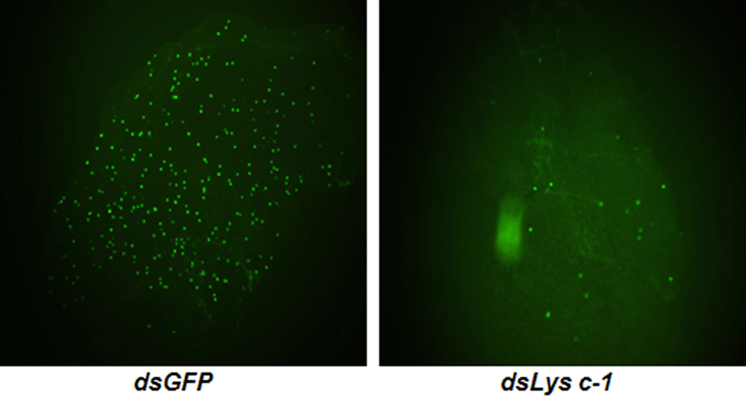
Fig. 6.
Comparison of An. dirus midguts infected with a GFP-expressing strain of P. berghei after the mosquitoes were injected with dsGFP or dsAdLys c-1. At 4 days after infection, midguts from each batch were dissected and the number of oocysts was counted using fluorescence microscopy. The number of parasites in dsAdLys c-1 KO mosquitoes was dramatically reduced when compared with control dsGFP mosquitoes.

