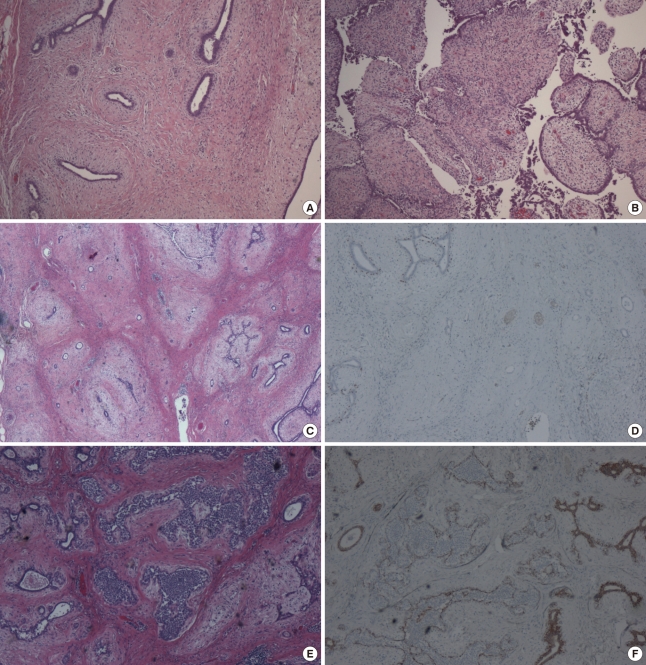
Figure 3.
(A) The mass revealed fibroadenomatous change (H&E stain, ×40). (B) Leaf-like structures of fibroepithelial lesion showed a cellular stroma with cytological atypia and increased mitosis (H&E stain, ×40). (C) Invasive carcinoma cells formed small tubules within the stroma (H&E stain, ×20). (D) Tubules of carcinoma cells were immunohistochemically negative for myoepithelial marker, p63 (IHC, ×40). (E) Lobular carcinoma in situ was noted within the glandular component (H&E stain, ×40). (F) Lobular carcinoma in situ was immunohistochemically negative for E-cadherin (×40).

